Triangular Fibrocartilage Complex Injury (TFCC)
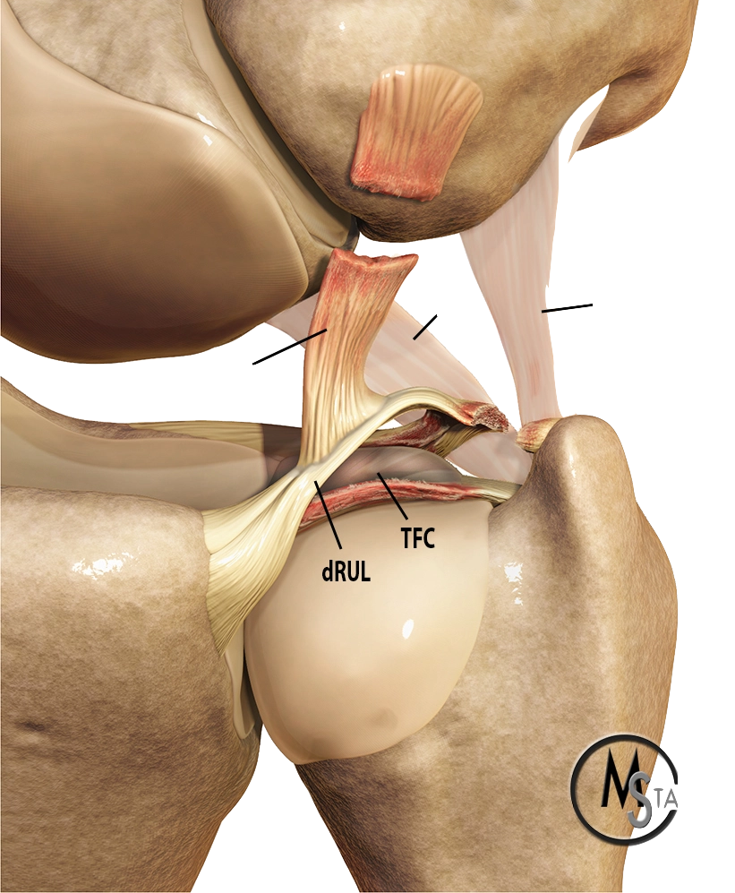
Triangular Fibrocartilage Complex Tear (TFCC)
The Triangular Fibrocartilage Complex (TFCC) is a complex structure that helps to stabilize and support the hand. It can be torn or injured as a result of repetitive use or an accident, leading to pain and inflammation. If you have suffered a TFCC tear, this guide will help you understand what it is, how it is treated, and what you can do to prevent future injuries.
Triangular Fibrocartilage Complex Tear (TFCC): An Introduction
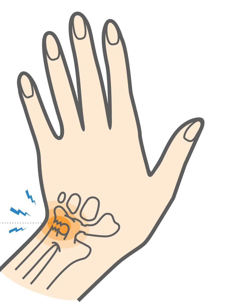
The triangular fibrocartilage complex (TFCC) is a group of structures located on the ulnar side of the wrist that helps to stabilize and cushion the joint. The TFCC can be injured by overuse or trauma, which can lead to pain and stiffness. A TFCC tear is a common injury in people who play sports or participate in other activities that involve repetitive wrist movements.
Treatment for a TFCC tear may include rest, ice, and physical therapy. Surgery may be necessary in some cases.
What Does the Triangular Fibrocartilage Complex Do?
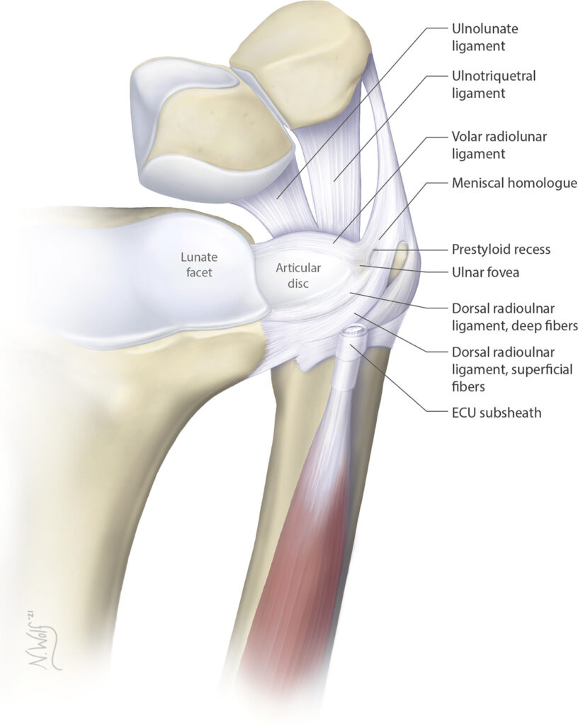
The triangular fibrocartilage complex (TFCC) is made up of several different structures, including:
* The articular disc: A rubbery pad that sits between the ulna and the carpus (the group of bones that make up the wrist)
* The ligaments: Strong bands of tissue that connect the bones and stabilize the joint
* The meniscus: A C-shaped piece of cartilage that helps to cushion the joint
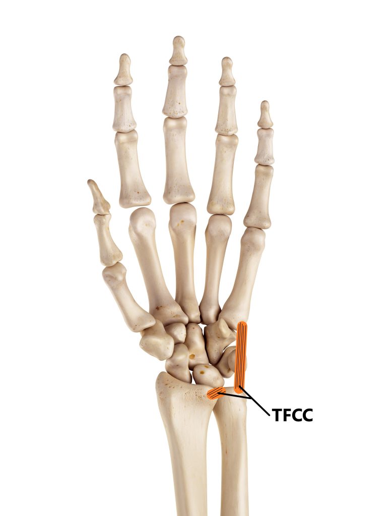
The TFCC is located on the ulnar side of the wrist (the side closest to the little finger). It acts as a shock absorber for the joint and helps to stabilize it during movement.
Symptoms of Triangular Fibrocartilage Complex Tear (TFCC)
The symptoms of a TFCC tear can vary depending on the severity of the injury. Some people may not have any symptoms, while others may experience pain, swelling, and weakness in the affected joint.
If you have a TFCC tear, you may notice:
– Pain in the wrist or forearm bones
– Swelling in the wrist
– Weakness in the affected hand or fingers
– Difficulty moving the affected joint
If you experience any of these symptoms, it is important to see a doctor so that they can properly diagnose and treat your condition.
Causes of Triangular Fibrocartilage Complex Tear (TFCC)
There are multiple causes of triangular fibrocartilage complex (TFCC) tears. The most common is chronic degenerative injury due to aging. Other causes include trauma, repetitive stress injuries, and certain medical conditions.
Degenerative wear and tear
Degenerative wear and tear is the main cause of TFCC tears in people over the age of 40. As we age, our bodies produce less collagen, a protein that gives our tissues their strength and elasticity. This decrease in collagen makes our tissues more vulnerable to injury.
Trauma
Trauma is another common cause of TFCC tears. A fall or direct blow to the wrist can tear the ligaments and cartilage that make up the TFCC. Repetitive stress injuries, such as those that occur from repetitive motions of the wrist (e.g., typing, weightlifting), can also lead to TFCC tears.
Certain medical conditions, such as arthritis and gout, can increase the risk of TFCC tears. People with these conditions often have inflammation in their joints, which can weaken the volar distal radioulnar ligaments and cartilage.
If you think you have a TFCC tear, it's important to see a doctor for an accurate diagnosis. Early diagnosis and treatment are essential for preventing further damage to the joint.
Diagnosis of Triangular Fibrocartilage Complex Injury
The diagnosis of a triangular fibrocartilage complex (TFCC) tear can be difficult. The first option to diagnose a TFCC tear is to have an X-ray of the wrist. An X-ray can show if there are any bony changes around the wrist joint that could be causing pain. However, an X-ray will not show a TFCC tear.
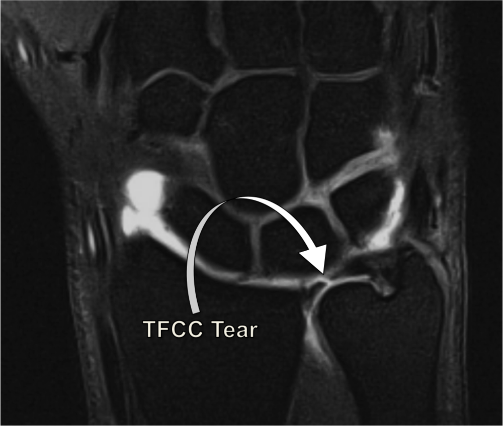
If your doctor suspects you have a TFCC tear, they may order an MRI of the wrist. An MRI can show the soft tissue structures, including the TFCC. An MRI is the best test to diagnose a TFCC tear.
Your doctor may also order a CT scan of the wrist. A CT scan can show the bones and soft tissues, but it is not as good as an MRI for diagnosing a TFCC tear.
Treatment for Triangular Fibrocartilage Complex Tear (TFCC)
Triangular fibrocartilage complex (TFCC) tears are a common injury, particularly in people who play sports or who have jobs that involve repetitive motion of the wrist. Treatment for a TFCC tear depends on the severity of the injury.
Mild TFCC Tears
If you have a mild TFCC tear, your doctor may recommend that you rest your wrist and avoid activities that aggravate your symptoms. You may also be advised to wear a splint or brace to immobilize your wrist and allow the TFCC to heal.
Moderate to Severe TFCC Tears
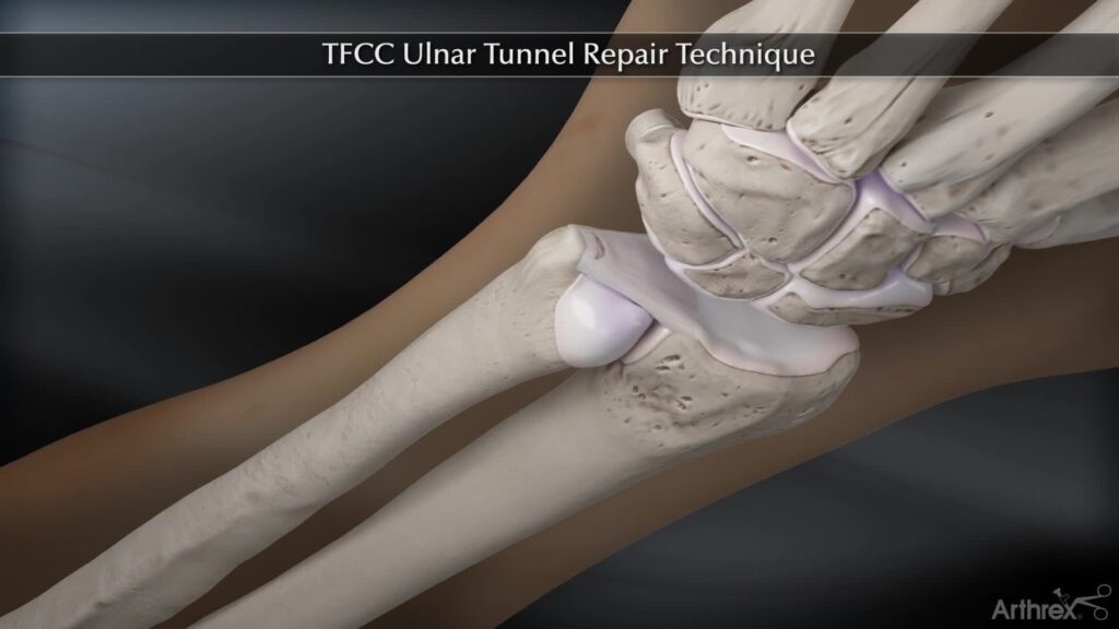
If you have a moderate to severe TFCC injury, your doctor may recommend surgery to repair the injury. Surgery is typically performed using an arthroscope, a small camera that is inserted into the joint through a small incision. The surgeon will then make additional incisions to insert surgical instruments.
During surgery, the damaged portions of the TFCC are removed and the remaining tissue is repaired. In some cases, a graft may be used to replace the damaged tissue.
After surgery, you will likely need to wear a splint or brace for several weeks. Physical therapy may also be recommended to help restore range of motion and strength to your wrist.
Outlook for patients with TFCC
The outlook for patients with Triangular Fibrocartilage Complex Tear (TFCC) largely depends on the severity of their condition. For those with a mild tear, conservative treatment such as to undergo physical therapy and splinting may be all that is necessary to improve symptoms. More severe or chronic TFCC tears may require surgery to repair the damage. In some cases, patients may need to have the entire TFCC removed.
The prognosis following surgery is generally good, but patients may experience stiffness and weakness in the affected hand.
Tips to prevent Triangular Fibrocartilage Complex Tear (TFCC)
Following are some of the ways to prevent TFCC injuries:
-Wear protective gear while participating in activities that may put you at risk for an injury to the TFCC, such as contact sports.
-Avoid repetitive motion of the wrist, such as when using a power drill or playing tennis.
-Use proper form and technique when lifting objects to avoid stressing the TFCC.
-Maintain a healthy weight to reduce the risk of developing osteoarthritis, which can contribute to the development of a TFCC tear.
If you have had a previous injury to the TFCC, be sure to follow your doctor’s recommendations for activity and exercise to help prevent further injury.
Frequently Asked Questions about Triangular Fibrocartilage Complex Tear (TFCC)
The symptoms of a TFCC tear can vary depending on the severity of the injury. However, common symptoms include ulnar sided wrist pain, weakness in the grip, swelling, and clicking or popping sensations when moving the wrist.
There are a variety of reasons why someone may develop an ulnar styloid impingement syndrome. Injuries caused by falls or other trauma to the wrist are one cause. Other factors that may contribute to developing an ulnar carpal impingement syndrome include overuse injuries, flexor muscle tendonitis, degenerative changes due to aging, or anatomical abnormalities.
TFCC tears are typically diagnosed with a combination of a physical examination and imaging tests. Your doctor will ask about your symptoms and medical history, and then perform a physical examination of your wrist. Imaging tests, such as X-rays, MRI, or CT scans, may also be ordered to help confirm the diagnosis.
The most common structures injured in a medial ankle sprain are the ligaments on the inside of the ankle near medial malleolus. These medial ligaments include the anterior talofibular ligament (ATFL), the calcaneofibular ligament (CFL), and the posterior talofibular ligament (PTFL). The ATFL is the most commonly injured ligament, followed by the CFL. The PTFL is the least common deltoid ligament injury.
The outlook for patients with a TFCC tear largely depends on the severity of their condition. For those with a mild tear, conservative treatment may be all that is necessary to improve symptoms. More severe tears may require surgery to repair the damage. In some cases, patients may need to have the entire TFCC removed. The prognosis following surgery is generally good, but patients may experience stiffness and weakness in the affected hand.
3 Ways to Level Up Your Rehab and Injury Prevention With Us


